Cherry Angioma
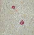
Cherry angioma is a common type of skin lesion that first occurs in early adulthood and continues with age. These round lesions typically develop on the trunk, hands, and feet; hey range from bright to dark red and can be up to several millimeters wide. Cherry angiomas are asymptomatic and have no reported clinical consequences.
The exact cause of cherry angioma is unknown but may be related to exposure to certain chemicals or changes in hormones. Treatment options for cherry angiomas include laser treatment, electrodesiccation for typical lesions, and surgical excision for larger lesions.
Sebaceous Hyperplasia (Senile Hyperplasia)
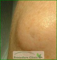
Sebaceous hyperplasia is a type of skin lesion common in middle-aged and elderly individuals. The lesions are soft, yellow, dome-shaped swellings that typically develop on the forehead, cheeks, and nose, and sometimes on the genitals. They usually range from 2 to 5 millimeters wide and are not harmful. Sebaceous hyperplasia can be confused with early basal cell carcinoma.
Treatment options include electrodesiccation, laser therapy, oral medication, and topical medication. Surgical excision can lead to scarring and is generally not considered. A skin biopsy may be done to check for basal cell carcinoma.
Seborrheic Keratosis
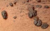
Seborrheic keratoses are thickened lesions on the skin that range from tan to brown to black with a clear border. The lesions typically have a rough surface and are 2 to 3 millimeters wide. They usually develop on the trunk but can also occur on the hands, feet, face, and scalp. Seborrheic keratoses increase in number as a person ages.
Seborrheic keratoses are not considered harmful but can become irritated and inflamed if rubbed or scratched. Treatment options include cryosurgery (freezing cells), curettage (surgical scraping), and surgical shaving excision. Topical steroids can be used on irritated lesions for symptomatic relief. A skin biopsy may be done to check for melanoma.
Dermatofibroma
Dermatofibromas are firm, raised nodules that develop on the body, most commonly on the lower legs. They range from brown to purple to yellow-pink and are usually 3 to 10 millimeters wide. The lesions are easily distinguished by their dimpling when pressed upon. It is unclear whether dermatofibromas are tumors, or reactions to minor trauma, insect bites, viral infections, ruptured cysts, or folliculitis.
Because dermatofibromas are located beneath the skin rather than on the surface, the ideal treatment option is surgical excision, rather than simply shaving the lesion.
Skin Tags
Skin tags are harmless growths that usually develop in the neck and underarm areas. They occur in 25 percent of the population and increase in number with age. Skin tags range in size from less than 1 millimeter to 1 centimeter wide and are typically a brownish color. They often occur in obese and overweight individuals, as skin friction leads to higher chance of skin tag development.
Simple excision can be used to remove skin tags, and cryotherapy is effective for smaller lesions. However, recurrences of skin tags are common. Skin tags are not usually signs of cancer, unless the patient has suffered symptoms since childhood, in which case they should be tested for basal cell carcinoma.
Keratoacanthoma
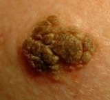
Keratoacanthomas are rapidly growing lesions that usually develop on sun-exposed skin in older individuals. They are often found on the face and limbs and occur more frequently in women. They can grow to become more than 2 centimeters wide in just two to four weeks. After a few months, the lesion rolls inward and leaves a hypopigmented (loss of skin color) scar. The exact cause of keratoacanthomas is unknown, but possible factors that contribute to the lesions include ultraviolet radiation exposure, human papillomavirus, and prolonged contact with coal tar derivatives.
Surgical excision is the preferred treatment method. Electrodesiccation, curettage (surgical scraping), or blunt dissection can be used for smaller lesions. Mohs’ surgery can be used in difficult areas, such as around the nose and ears.
Pyogenic Granuloma
.JPG)
Pyogenic granulomas are red or yellow bumps that easily bleed. They often develop on the head, neck, arms, and legs, starting off with a small lesion but growing quickly. The exact cause is unknown, but viruses, trauma, and burns may play a role.
Although pyogenic granulomas sometimes roll inward within six months, most patients seek treatment because of bleeding. Treatment options include surgical excision (with option of laser therapy), or shave excision with curettage (surgical scraping) and cautery (use of heat). The lesions may recur if not removed completely.
Epidermoid Cyst
Epidermoid cysts, also known as inclusion cysts and epidermal inclusion cysts, are round capsules ranging in size from a few millimeters to several centimeters. They usually occur on the face, back, and chest. The cysts may remain small for years or may grow rapidly, and rupture of a cyst initiates an inflammatory response. The pus can either drain from the surface or be slowly resorbed.
Surgical excision is the preferred treatment method. Some cysts may need to be drained prior to removal. Sutures may be needed for larger cyst removal.